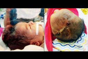
The huge 10-cm swelling in the baby's head went undetected in scans till her birth; doctors at the Surya hospital in Santacruz begin research to understand the growth

The baby was born with a huge growth on her scalp, which was later removed by surgeons
An infant born in the city with a rare, huge lesion growth in her head has inspired neurosurgeons who treated her to pore over research books to find out the exact reason behind the enlargement, which they've fortunately found to be non-cancerous.
ADVERTISEMENT
The case has developed great interest amidst doctors because the growth of the foetus was being monitored by a gynaecologist and nephrologist [since the mother was undergoing renal treatment]. All ultra sonography (USG) reports showed normal growth of the foetus. The last sonography was done 20 days before the delivery and experts feel something happened during this period, leading to such a huge growth on the baby's scalp. The baby was born on February 25.
The case
Dr Shraddha Maheshwari, consulting neurosurgeon, who saw the child at Surya hospital on the day she was born said, "The child was born in a nursing home in Malad and everything was normal, apart from a huge lesion growth in the centre of the head. She was referred to Surya hospital in Santarcruz west. Upon seeing her, I was shocked to find out about the lesion, which measured almost 8 cm by 10 cm in circumference, with a height of 4 cm."

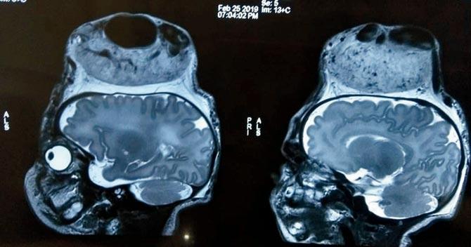
An MRI scan of the baby's head revealed that the brain was normal, but there were a few abnormal blood vessels passing blood to the lesion
During an initial briefing from the family, the doctors learnt that the newborn's mother was undergoing treatment for a renal problem. At Surya hospital, the team of doctors and consulting neurosurgeons were concerned about the diagnosis and line of treatment. Having no prior diagnosis papers in hand, they decided to get an MRI scan done.
"During the physical examination, we initially suspected it to be a case of either a brain tumour or a cancerous growth. We also suspected that a portion of the brain might have protruded from her scalp. Some fluid was slowing dripping from an opening on the lesion, too, which was not a good sign," explained Dr Maheshwari, adding, "Luckily, the MRI scan showed us that the brain was normal, but there were a few abnormal blood vessels passing blood to the lesion through the scalp."
On February 26, the baby underwent supra major surgery, which lasted for over four hours. Dr Shashank Joshi, another consulting neurosurgeon, who was also a part of the operating team said, "Unlike adults, who usually have five to six litres of blood in their body and can withstand any surgery, newborns usually have around a litre of blood in their body. Operating on the infant in this case was a bit challenging as there were seven to eight blood vessels supplying blood to the lesion, and even a small amount of blood loss could have been fatal."
Dr Joshi added, "We had to be very careful because the blood vessels were highly dilated, tortuous and expanded. However, the infant was able to withstand the procedure and required only a single bag of blood transfusion."
Dr Maheshwari added, "Post the removal of the lesion, instead of skin grafting, we decided to use the skin on the baby's scalp to cover the opening. We did the V-Y plasty to stitch the wounds. The child was put on ventilator support. Post surgery, around 36 hours were crucial." The baby responded well to the treatment in the neonatal care unit.
Despite the successful surgery, worries still persisted in the minds of the surgeons and doctors until the return of the lesion sample sent to the Tata Hospital. On March 7, the Tata hospital sent its report (mid-day has a copy) ruling out the lesion from being cancerous. Instead, it said that the growth was a hemangioma, which is a benign tumour made up of blood vessels.
Dr Nandakishor Kabra, neonatologist and director of neonatal intensive care at Surya child care hospital, under whose care the newborn was post surgery, said, "This is one of the most unique cases I've come across in my entire career. The initial two days of post operative care for the baby were a bit challenging as we had to closely monitor the neurological behaviour of the infant to ensure the oxygen level and cardiac parameters were maintained. There was a lot of ambiguity about the growth, which luckily turned out to be non-cancerous in the histopathology findings. The baby is doing quite well." The newborn's father Biru Malli is happy too, "We are thankful to the surgeons and doctors who relieved my child from the lesion. We're happy to have her home."
In international journals
Now, the infant's unique condition will make its way to international medical journals. The neurosurgical team of Dr Maheshwari and Dr Joshi are in the process of writing papers on their experience in this case and will be submitting the same for publication. Prior to that, they are also doing further research to find the source where the lesion was developed, since it was not visible in the USG report.
Expert says
Explaining hemangioma, Dr Y S Nandanwar, professor at DY Patil medical college said, "Hemangioma is a rarest of the rare case, and can be detected in the womb in a USG, only if someone is specifically looking for minute vascular anomalies in the foetus. Usually, such anomalies are looked for in the initial few months of pregnancy, because there are fewer chances of spotting any abnormal growth post five months as the position of the foetus's head is downwards, falling under the mother's pelvic shadow." Asked about the probable chances of abnormal growth between the last sonography and the delivery, Dr Nandanwar responded in the affirmative, stating, "Since hemangioma is an abnormal development of blood vessels, such lesions can occur anytime. This is more commonly seen with female foetuses. Medical science is yet to find the reason for such an abnormal growth of blood vessels. Early diagnosis and proper treatment are crucial."
Catch up on all the latest Crime, National, International and Hatke news here. Also download the new mid-day Android and iOS apps to get latest updates
 Subscribe today by clicking the link and stay updated with the latest news!" Click here!


